This post was first published in August 2022
Updated on 17th September 2025

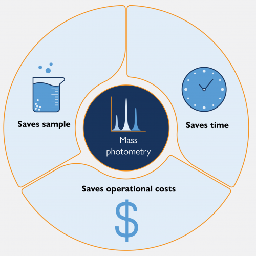
Mass photometry offers significant savings in terms of sample, user and instrument time, and operational costs.
For structural biology studies, it is key to conduct a proper assessment of samples prior to cryo-EM measurements – as sample preparation is not always straightforward, and instrument time is too precious to waste on subpar samples.
Over the last few years, mass photometry has started to enter more and more laboratories working within the structural biology space. This is a result of the recognition of the time- and cost-saving benefits to be had from utilizing mass photometry as a quick and experimentally straightforward method for screening sample quality prior to cryo-EM analysis.
Mass photometry requires minimal sample and can measure biomolecules in their native state, with its single-molecule resolution over a wide dynamic range making it ideally suited for assessing sample monodispersity. In contrast with other techniques like negative stain EM (NS-EM), mass photometry has low turnaround times and operating costs – which streamlines the analytical process. Below, we answer frequently asked questions about what mass photometry can do for structural researchers working with cryo-EM.
“Compared with other biophysical approaches, including SEC and DLS, MP experiments require lower sample amounts (10–20 μL) and are amenable to lower (nM range) concentrations of protein. This allows for testing of several conditions prior to cryo-EM without depleting all or most of the sample. Finally, MP is a straightforward technique even for novice users. The instrument is set up in an existing lab space, and the user-friendly software does not require a highly trained operator. Together, these factors establish MP as a powerful tool for evaluating sample quality prior to structural studies.”
Mass photometry measures the mass of single particles in solution and uses a histogram to report the mass distribution of the sample, providing an overview of all the populations in a sample. You can read more about how mass photometry works here and learn more about interpreting mass photometry data here.
Mass photometry helps determine sample purity and measure the percentage of a sample population that has formed a desired complex before proceeding with cryo-EM. Collecting these data is an important step in sample quality control that can help you improve structural resolution and data reproducibility. Mass photometry has been shown to be an effective method for measuring aggregation and oligomerization [1]. Others have found that the information about sample composition not only is comparable to that provided with NS-EM, but also able to detect components in the sample invisible to NS-EM [2].
Mass photometry has been shown to agree well with data from negative stain EM, charge detection mass spectrometry (CD-MS) and native MS, as described in [2]–[4]. The advantage of mass photometry over these techniques is that it is much faster and simpler to use.
In contrast to light-scattering techniques, like dynamic light scattering (DLS) or multi-angle light scattering (MALS), mass photometry is a single-particle technique. This means that it counts how many particles in your sample have a particular mass. Coupled with its wide dynamic range and wide mass range, this makes mass photometry more sensitive to the detection of low abundance species, such as aggregates, and provides access to sub-populations within samples (for more on the differences between DLS, MALS and mass photometry, watch this webinar).
When compared to other commonly used techniques, mass photometry has shorter measurement times and consumes less sample.
Each mass photometry measurement takes only one minute. As one mass photometry user from a structural biology lab told us, “Typically, I can check the sample in one minute by mass photometry, whereas my negative stain would probably have taken a couple of hours.” This is a very powerful advantage of mass photometry, which can save hours in the optimization phase of experiments [5].
Mass photometry does not require specific buffers; it is compatible with the most widely used buffers. This wide compatibility, along with its low time and sample requirement, make mass photometry ideal for repeated testing to optimize buffer conditions.
Mass photometry is also compatible with detergents and other membrane mimetics, so you can often keep membrane proteins – as well as biomolecules that are soluble in aqueous media – in solution during analysis. That way, it is possible to keep biomolecules in their native states, or as close to them as possible.
Mass photometry is a label-free technique that measures the mass of biomolecules in their native state. There is no need for the hazardous heavy metals used in negative stain EM or for drying out samples, which can introduce artifacts in the data and make it more difficult to optimize buffer conditions later for cryo-EM.
Mass photometry measurements require very little sample – about 10 – 20 µL. The optimum sample concentration is typically at the low nanomolar level. There will be plenty of sample left over to use for cryo-EM after a mass photometry experiment.
For mass photometry measurements of proteins in the context of structural biology, Refeyn offers the TwoMP mass photometer, along with all the necessary consumables. If repeated analysis of large sets of sample is needed, there is an automated version of the TwoMP. For the analysis of low-affinity interactions that require higher concentrations, a microfluidics add-on – MassFluidix™ HC – is also available.
The mass range of the TwoMP instrument is 30 kDA to 5 MDa, and this range can vary depending on experimental conditions, such as the sample carrier slides used. Refeyn’s MassGlass™UC sample carrier slides can handle 50 kDa to 5 MDa out of the box, with the possibility of lowering the mass range to the 30 kDa minimum after cleaning them by hand.
The TwoMP is a low-footprint instrument that fits on a benchtop. It does not need its own dedicated room, unlike an electron microscope. The TwoMP comes with its own anti-vibration device, and the entire setup can fit neatly into most laboratories without additional construction.
Blog – Enhancing cryo-EM workflows with mass photometry: A smarter pre-screening approach – Refeyn
If you want to read more about how mass photometry supports cryo-EM studies, this blog post contains detailed explanations, as well as case studies from the literature that highlight how researchers over the world are integrating mass photometry into their research.
Webinar: Unlocking Complex Sample Analysis: How Mass Photometry Simplifies Protein Characterization
This webinar features Perla Vega, Technical Sales Specialist at Refeyn, as she explains the key principles and applications of mass photometry, from antibody aggregation to protein-DNA interactions. Later, Dr. Philip Kitchen from Aston University shares real-world case studies showcasing how mass photometry helps tackle the challenges of membrane protein analysis. Learn how this technique quickly and easily characterizes protein purity, interactions, and oligomerization, making it a valuable tool for research and biopharma applications.
Read more about the TwoMP mass photometer
In this product data sheet, we provide details on the specifications and features of the TwoMP and the easy-to-use software that comes with it.
How confident are you in the quality of your cryo-EM sample?
[1] S. Niebling et al., “Biophysical Screening Pipeline for Cryo-EM Grid Preparation of Membrane Proteins,” Frontiers in Molecular Biosciences, vol. 9, p. 535, Jun. 2022, https://doi.org/10.3389/fmolb.2022.882288
[2] A. Sonn-Segev et al., “Quantifying the heterogeneity of macromolecular machines by mass photometry,” Nature Communications, vol. 11, no. 1, pp. 1–10, Dec. 2020, https://doi.org/10.1038/s41467-020-15642-w
[3] V. Yin et al., “Probing Affinity, Avidity, Anticooperativity, and Competition in Antibody and Receptor Binding to the SARS-CoV-2 Spike by Single Particle Mass Analyses,” ACS Central Science, vol. 7, no. 11, pp. 1863–1873, Nov. 2021, https://doi.org/10.1021/acscentsci.1c00804
[4] M. A. den Boer, S.-H. Lai, X. Xue, M. D. van Kampen, B. Bleijlevens, and A. J. R. Heck, “Comparative Analysis of Antibodies and Heavily Glycosylated Macromolecular Immune Complexes by Size-Exclusion Chromatography Multi-Angle Light Scattering, Native Charge Detection Mass Spectrometry, and Mass Photometry,” Analytical Chemistry, vol. 94, no. 2, pp. 892–900, Dec. 2021, https://doi.org/ 10.1021/acs.analchem.1c03656
[5] Y. Xu and S. Dang, “Recent Technical Advances in Sample Preparation for Single-Particle Cryo-EM,” Frontiers in Molecular Biosciences, vol. 9, p. 892459, Jun. 2022, https://doi.org/10.3389/fmolb.2022.892459