- TwoMP
-

For molecular mass measurements with unmatched sensitivity, speed and simplicity of use, a TwoMP mass photometer offers a wide mass range and single-molecule resolution.
High-fidelity measurements of molecular mass
Little sample required
Intuitive acquisition and data analysis software
Easy setup – a compact, benchtop instrument with minimal installation requirements
- TwoMP Auto
-
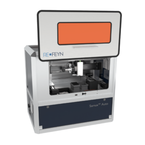
The automated mass photometer frees up operator time and offers enhanced precision, enabling rapid measurement of multiple samples with low sample consumption.
Rapid, automated measurement of multiple samples
Ideal for screening and titration assays
Highly reproducible data with user-friendly software
- MassFluidix HC
-
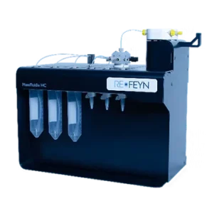
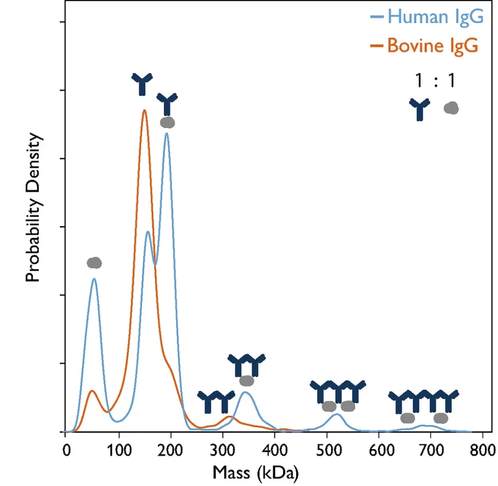
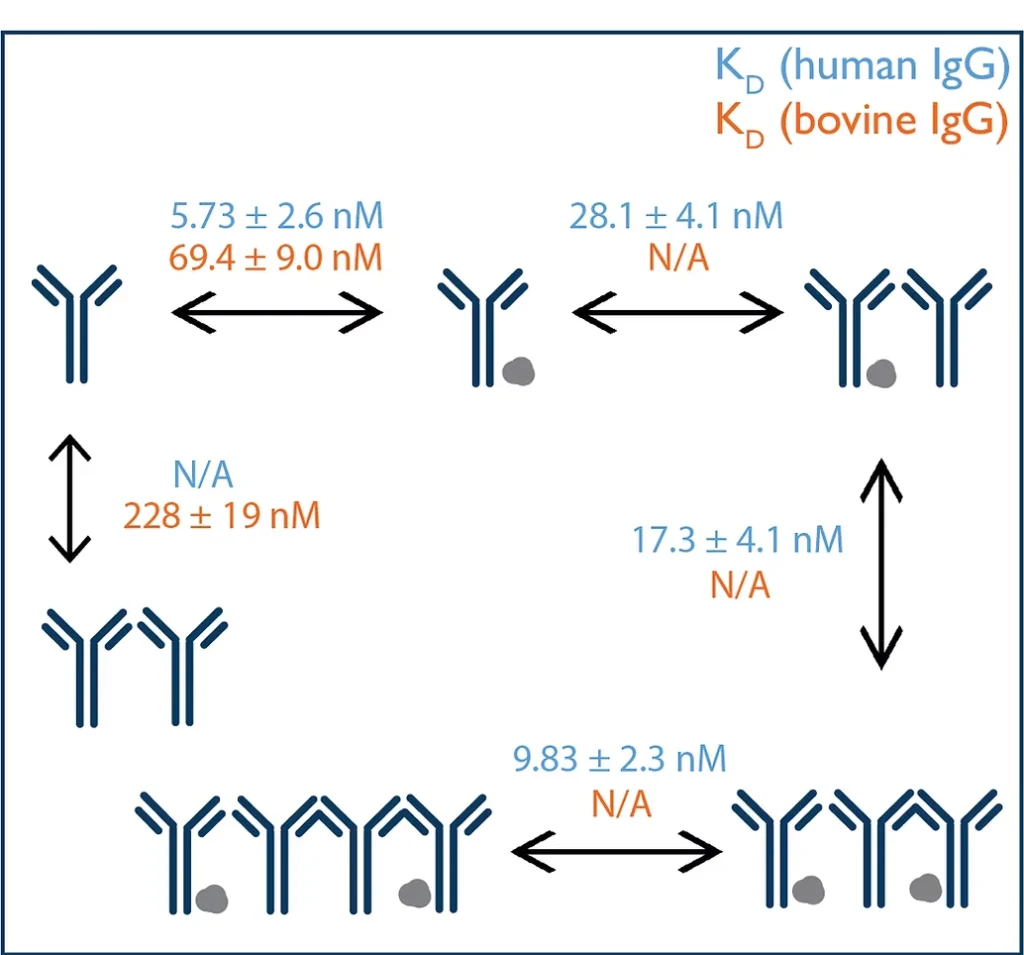
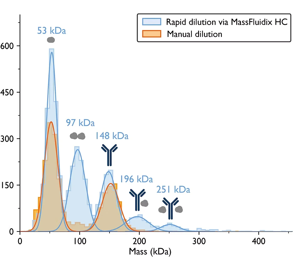
Refeyn’s microfluidics system, MassFluidix HC, significantly expands the range of sample concentrations amenable to investigation by mass photometry, by raising the upper sample concentration limit from the nanomolar to the micromolar range. This enables applications such as the characterization of low-affinity interactions.
- Enables measurement of concentrated samples
- Uses rapid dilution
- Is an add-on for TwoMP and OneMP mass photometers
- Refeyn consumables
-
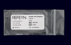 By using Refeyn consumables, you can spend less time preparing for measurements and gain greater confidence in your data. The consumables range includes calibrants, sample carrier slides and more.
For molecular mass measurements with unmatched sensitivity, speed and simplicity of use, a TwoMP mass photometer offers a wide mass range and single-molecule resolution.
High-fidelity measurements of molecular mass
Little sample required
Intuitive acquisition and data analysis software
Easy setup – a compact, benchtop instrument with minimal installation requirements
![]()
The automated mass photometer frees up operator time and offers enhanced precision, enabling rapid measurement of multiple samples with low sample consumption.
Rapid, automated measurement of multiple samples
Ideal for screening and titration assays
Highly reproducible data with user-friendly software
Refeyn’s microfluidics system, MassFluidix HC, significantly expands the range of sample concentrations amenable to investigation by mass photometry, by raising the upper sample concentration limit from the nanomolar to the micromolar range. This enables applications such as the characterization of low-affinity interactions.
- Enables measurement of concentrated samples
- Uses rapid dilution
- Is an add-on for TwoMP and OneMP mass photometers