Home/Mass photometry analysis of GPCRs in copolymer nanodiscs
Copolymer nanodiscs solubilize GPCRs
G-protein-coupled receptors (GPCRs) are integral membrane proteins that fulfill essential biological roles and are frequent therapeutic targets.
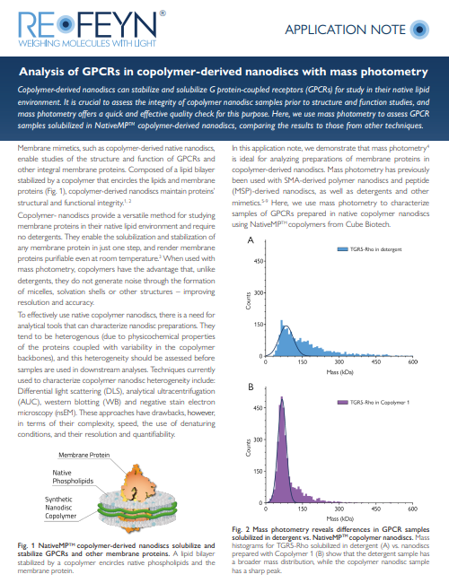
As with other membrane proteins, GPCRs contain hydrophobic domains that disrupt the protein’s structure and activity in aqueous buffer. For this reason, membrane mimetics are used to solubilize and stabilize membrane proteins.
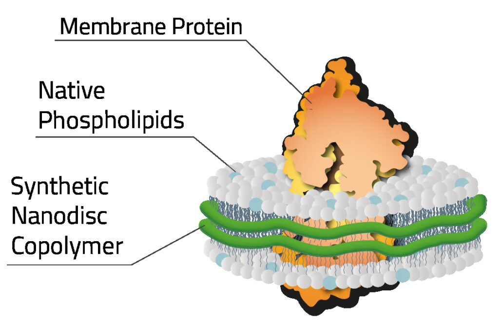
Composed of a lipid bilayer stabilized by a copolymer that encircles the lipids and membrane proteins, copolymer-derived nanodiscs enable the study of GPCRs and other membrane proteins without detergents.
Mass photometry characterizes nanodisc-solubilized GPCRs
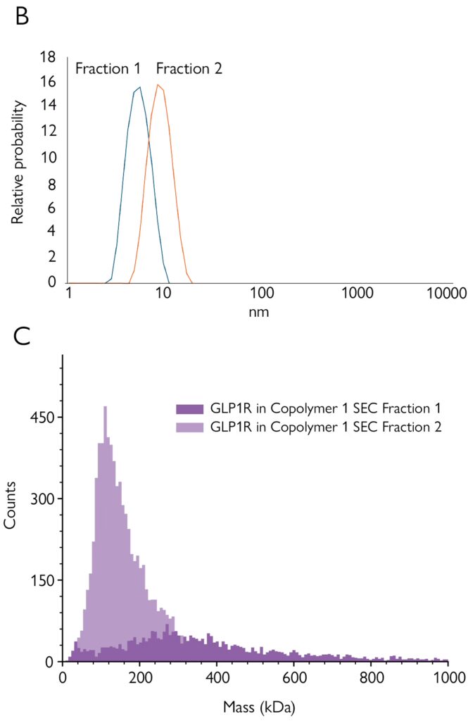
In this application note, created in collaboration with CUBE Biotech, copolymer-derived nanodiscs were used to solubilize GPCRs. The embedded proteins were fractionated using SEC, then characterized with SDS-PAGE, western blot, DLS and mass photometry.
Mass photometry characterized the SEC fractions quickly and with minimal sample preparation – providing a more detailed overview of the populations than other techniques. These advantages make mass photometry well-suited for efficient quality checks during membrane protein preparation prior to structural studies or functional assays.
Discover how mass photometry characterizes
nanodisc-embedded GPCRs
Analysis of GPCRs in copolymer-derived nanodiscs with
mass photometry
Discover how to use mass photometry to expedite functional assays or structural investigations by obtaining detailed information on your membrane protein samples or SEC fractions. Learn how copolymer-derived nanodiscs can be used for GPCR solubilization, and how mass photometry compares to other techniques in this context.